
Common Tests and Therapies
 Cardiac Catheterization
 Echocardiogram
 Echocardiograms
Like a sonogram, these procedures use sound waves from a transducer (wand) to bounce off your heart, which produce a reflection of your heart in motion. That reflection is captured in a video image, which can help detect structural problems in the heart. Echocardiograms can be conducted while at rest or while exercising (a stress echo). There are a few different types of these tests:
- Transthoracic Echocardiography (TTE) – TTEs use ultrasound to get a fuller picture of the heart’s size, structure, and motion. In this simple, painless procedure, you lie on your left side while a technician moves a device over your chest. The device collects images of your beating heart.
- Transesophageal Echocardiography (TEE) – TEE is often used before or during heart surgery to guide post-operative treatment or see if additional work is needed before you leave the operating room. A physician places a long tube with an ultrasound probe in your esophagus while you are under sedation. The probe creates an ultrasound “movie” of your heart at work, giving a much clearer picture than is possible with an electrocardiogram (below) alone.
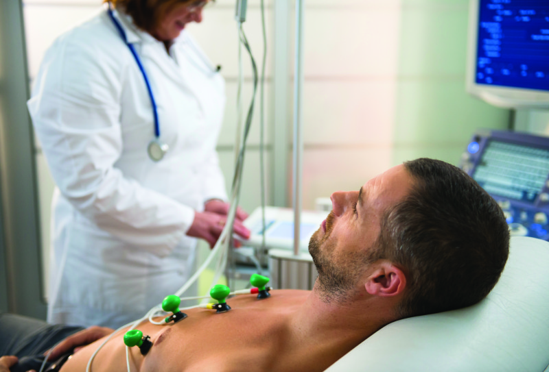
Electrocardiogram (ECG or EKG)
This test measures the heart’s electronic activity to assess its electrical output and see if there are problems.
What to Expect
This simple, painless procedure involves placing patches with electrodes to locations on your skin to measure electrical activity of the heart, either while you’re at rest or during activity. The activity is charted on a small screen or printed on paper. The test lets your doctor know how fast and steadily your heart is beating and the strength of the signals at each stage of your heart’s beating. The consensus is that this procedure is low risk for most patients.
Myocardial Perfusion Imaging (MPI)
MPI, also referred to as a nuclear stress test, takes pictures of the blood flow pattern to the heart muscle using a radioactive tracer and special imaging equipment. Nuclear medicine is a powerful way to catch diseases in their earliest stages by letting doctors see molecular changes in the body.
What to Expect
A radioactive tracer is swallowed, inhaled, or injected and in time gathers near the examination site. Imaging equipment picks up on the radioactive emissions from the tracer and provides a detailed image of the area. The consensus is that this procedure is low risk for most patients.
Positron Emission Tomography (PET) Scan
PET scans also use a radioactive tracer to identify issues with the body’s organs and tissues. A PET scan of the heart allows your doctor to assess if your heart is receiving enough blood, see heart damage or scar tissue, and observe buildup in the heart muscle.
What to Expect
A heart PET scan takes about 90 minutes and is done in a facility with a PET scanner. You’ll first receive a tracer by injection. It takes an hour to travel through your bloodstream and gather in your organs and tissues. Then you lie on a table that moves into a tunnel-shaped scanner, which picks up signals from the tracer and generates 3-D pictures of your heart.
PET scans are generally safe—the radiation use is similar to that of a CT scan, and it doesn’t stay in your body for long. Some experience pain or irritation at the injection site. Women who are pregnant or breastfeeding should let their doctor know.
Heart Monitors
Small, portable monitoring devices are also used to detect heart issues or see if heart treatments are working. Holter and event monitors are most often prescribed to diagnose and assess arrhythmias in your heart or detect silent cardiac ischemia (heart disease without symptoms). They can be worn under clothes while you perform normal activities. Here are the two main types:
- Holter Monitor – This one records all your heartbeats continuously. Electrodes are attached to your chest, and the monitor picks up the electrical activity of your heart at all times, even while sleeping.
- Event Monitor – Similar to a Holter monitor, this one records activities only at certain times.